Indirect immunofluorescence (IIF) is an important screening method for laboratory diagnostics due to its high sensitivity and specificity together with its broad antigenic spectrum. However, the microscopic evaluation of the fluorescence patterns is time-consuming and challenging for laboratory staff. Nowadays, many laboratories use automated systems to facilitate and standardise the IIF readout and interpretation. Automated microscopy systems enable fast digital acquisition of immunofluorescence images, as well as reliable result evaluation encompassing discrimination of positive and negative samples, pattern classification for key autoantibodies, and titer designations. New state-of-the-art systems incorporate artificial intelligence (AI) based on deep learning methods for classification of the immunofluorescence patterns and calculation of the antibody titer. The advent of live microscopy, whereby the evaluation is performed completely on-screen, provides a new level of speed and convenience for IIF diagnostics, as well as high standardisation between microscopes and operators. Automated IIF result interpretation is particularly useful for autoimmune serological applications such as detection of anti-nuclear antibodies (ANA) on human epithelial (HEp-2) cells, anti-neutrophil granulocyte cytoplasm antibodies (ANCA) on granulocytes, or anti-double-stranded DNA (dsDNA) antibodies on Crithidia luciliae.
ANA
ANA represent a key diagnostic criterion for many autoimmune diseases, especially rheumatic diseases such as systemic lupus erythematosus (SLE), mixed connective tissue disease, Sjögren’s syndrome, systemic sclerosis and poly/dermatomyositis. The gold standard for ANA determination is IIF on HEp-2 cells. This substrate provides the complete antigen spectrum and allows investigation of over 100 different autoantibodies. Positive results from IIF are confirmed using monospecific tests such as ELISA, chemiluminescence immunoassays (ChLIA), immunoblot or IIF microdot assays.
Related: Comprehensive analysis of SARS-CoV-2 immune responses
Identification of the fluorescence pattern on HEp-2 cells enables classification of the antibody or antibodies present in the patient sample. The International Consensus on Antinuclear Antibody (ANA) Patterns (ICAP, www.anapatterns.org) has developed a classification tree of patterns, with each assigned an anti-cell (AC) code number, to harmonise reporting between laboratories.
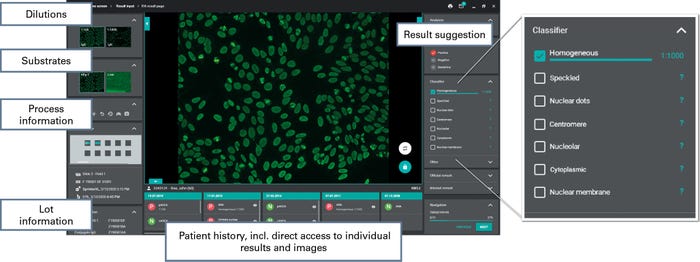
The evaluation of ANA on HEp-2 cells can be simplified and standardised using computer-aided microscopy systems, with those based on artificial intelligence providing the highest proficiency. The EUROPattern system, for example, uses deep convolutional neural networks to provide highly reliable differentiation of positive and negative ANA results, as well as identification of nine ANA patterns according to ICAP, namely homogeneous (AC-1), speckled (AC-4, 5 29), dense fine-speckled (AC-2), nucleolar (AC-8, 9, 10), nuclear dots (AC-6, 7), centromere (AC-3), nuclear envelope (AC-11, 12), anti-mitochondrial antibodies (AMA, AC-21), and cytoplasmic (AC-15 to 23). The deep learning algorithms ensure efficient segmentation of the HEp-2 cells, that is detection of their location and shape, so that counterstaining of the cells is no longer necessary. Both interface and mitotic cells are reliably identified. The automatic classifier generates pattern and titer suggestions with confidence values, including for mixed patterns, which occur where more than one antibody is present in the sample. For each pattern the titer is automatically calculated from the fluorescence intensities of the incubated dilutions, ensuring reproducible results.
Related: Euroimmun Webinar: Dermatomycoses – Diagnostic challenges
To investigate the diagnostic accuracy of the automated procedure, IIF evaluation using the EUROPattern classifier was compared to conventional visual interpretation using 213 patient sera. The overall agreement for positive/negative discrimination amounted to 93.0%. In the pattern assignment the agreement ranged from 90.1% to 100% for the different nuclear patterns, and 85.4% to 99.5% for the cytoplasmic patterns.
Anti-dsDNA antibodies
Anti-dsDNA antibodies are a hallmark of SLE and represent an important criterion for diagnosis. Their prevalence in SLE ranges from 20% to 90% in different studies, depending among other things on the test method used and the disease activity. Like the gold standard Farr assay, IIF using Crithidia luciliae as the substrate (CLIFT) is considered to have a very high disease specificity. The method takes advantage of the kinetoplast of Crithidia luciliae, which is rich in DNA but contains hardly any other antigens, thus enabling highly selective detection of anti-dsDNA antibodies. Automated evaluation of the fluorescence signals increases the reliability of the results compared to manual reading, which is subjective and leads to high intra- and inter-laboratory variation.
Interpretation of CLIFT is incorporated into the EUROPattern system. The sophisticated software is able to recognise the organelles of the protozoan and evaluates the specific kinetoplast fluorescence rather than just dark-light classification, ensuring high result reliability. Results are classified as positive or negative depending on the kinetoplast fluorescence, and include a titer designation based on the fluorescence intensity for positive samples. In a comparison of automated and visual evaluation of Crithidia luciliae IIF using 83 sera, the agreement between the two methods was 97.6%.
ANCA
ANCA are important serological markers for diagnosis and differentiation of autoimmune vasculitides, especially granulomatosis with polyangiitis (GPA, formally known as Wegener’s granulomatosis), which is characterised by autoantibodies against proteinase 3 (PR3), and microscopic polyangiitis (MPA), which is typified by autoantibodies against myeloperoxidase (MPO). In addition, ANCA can be found in chronic inflammatory bowel diseases. ANCA are detected by IIF with confirmation using monospecific assays.
The IIF substrates ethanol-fixed and formalin-fixed granulocytes are used to identify the typical ANCA staining patterns of anti-PR3 (cytoplasmic, cANCA) and anti-MPO (perinuclear, pANCA) antibodies. An additional substrate consisting of HEp-2 cells coated with granulocytes allows immediate differentiation between ANCA and ANA. The EUROPattern system incorporates automated positive/negative identification of ANCA, as well as pattern recognition for pANCA, cANCA and atypical ANCA (DNA-ANCA, xANCA). The latter can arise from antibodies against lactoferrin or other antigens. An estimated titer with a confidence value is given.
The computer-aided evaluation of ANCA with EUROPattern was compared to manual evaluation using 170 sera incubated on a BIOCHIP mosaic of ethanol-fixed granulocytes and formalin-fixed granulocytes. The overall agreement of results amounted to 98.2%. For the pattern assignment there was an agreement of 96.5% for cANCA, 94.7% for pANCA and 91.8% for atypical ANCA.
Further autoantibodies
The determination of autoantibodies on tissue substrates facilitates diagnosis of a range of autoimmune diseases. For example, investigation of autoantibodies in autoimmune liver diseases using the substrates rat liver and rat kidney supports the diagnosis and differentiation of autoimmune hepatitis type 1 and 2 and primary biliary cholangitis. Immunofluorescence signals on these two substrates can be evaluated automatically with the EUROPattern system. The evaluation includes positive-negative classification for relevant ANA (rat liver) and AMA (rat kidney), as well identification of an anti-liver kidney microsome (LKM) pattern using both substrates for reciprocal confirmation. The agreement between automated and visual evaluation on the tissue substrates amounted to 96.5% for ANA and 98.6% for LKM on rat liver and 94.6% for AMA and 98.9% for LKM on rat kidney.
Automated image acquisition for further tissue substrates, such as monkey liver, monkey stomach and monkey oesophagus, as well as for cell-based assays is also available.
Live and automatic microscopy
On-screen microscopy simplifies the IIF evaluation immensely by allowing recording and viewing of IIF images directly at the computer screen, thus eliminating the need for a dark chamber. The new EUROPattern Microscope Live (Figure 1) combined with EUROLabOffice 4.0 software (Figure 2), in particular, provides state-of-the-art live microscopy and fully automated acquisition of fluorescence images. The novel automatic laser focussing enables fast image acquisition and classification within two seconds per image. The images are recorded by means of a high-resolution camera, generating high-definition pictures. The microscope includes a self-calibrating long-life fluorescence LED which provides constant illumination, ensuring standardised quality between microscopes even at different locations. For live microscopy the multi-touch screen of the monitor allows easy navigation and zooming. Team consultations can be undertaken without the need for a discussion bridge.

Figure 1: EUROPattern Microscope Live
The EUROLabOffice 4.0 software classifies the fluorescence patterns and designates titers as described above. It communicates continuously between the LIS, the automatic processor and the microscope, ensuring rapid, secure, and traceable data exchange. Its complete network integration means that IIF results can be consolidated with findings from other analysis methods such as ELISA, immunoblot or ChLIA to produce a detailed and substantiated patient report. All information is presented clearly in the results window. Findings from different serum dilutions and substrates are consolidated into one report per patient, and new results are compared with the patient’s history. Negative samples can be confirmed in batches for added efficiency. All data, results and images are archived without the need for paper records.

Figure 2: Example of ANA evaluation with EUROPattern
Perspectives
Computer-aided evaluation provides standardisation and consolidation of IIF results in autoimmune diagnostics. Automation platforms with harmonised software and hardware reduce workload for laboratory technicians and provide a more objective evaluation. The progression from hands-on microscopy to complete on-screen evaluation has resulted in an unprecedented level of convenience and efficiency in IIF diagnostics. Moreover, software incorporating deep learning processes provides accurate positive/negative classification, pattern recognition and titer designation at a quality equivalent to or better than visual microscopy. Further applications with pattern evaluation using neural networks will soon be added to the repertoire.
This article appears in the latest issue of Omnia Health Magazine. Read the full issue online today.


.webp?width=700&auto=webp&quality=80&disable=upscale)